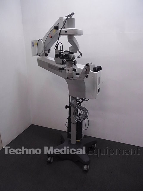
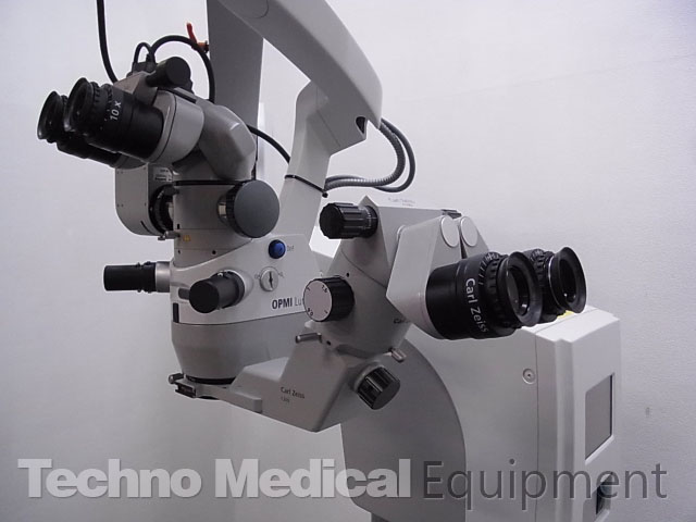
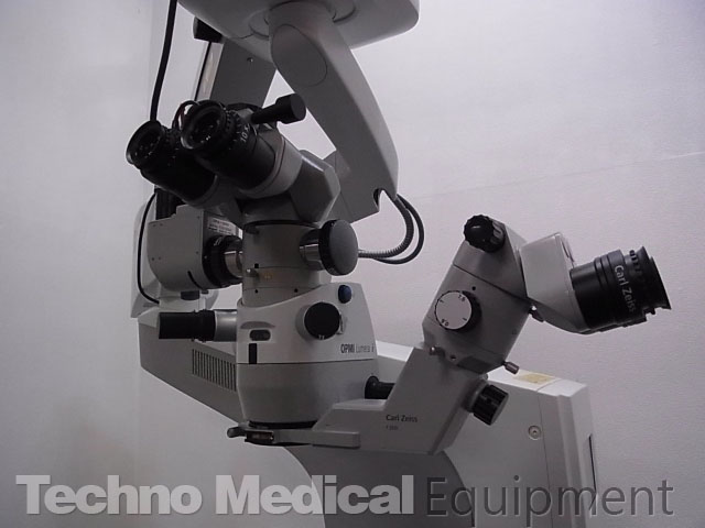
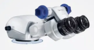
Carl Zeiss OPMI Lumera i Surgical Microscope

Click to learn more
Carl Zeiss OPMI Lumera i Surgical Microscope, Very good condition, patient ready
Patient ready condition, the optical system is very clear and perfect condition
Manufacture year: 2013, All items are sold in as-is condition with 6 month warranty.
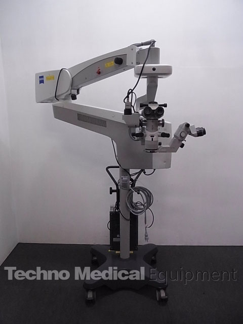
Carl Zeiss OPMI Lumera i Surgical Microscope includes:
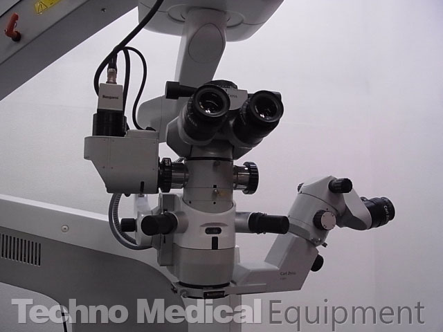
- Zeiss OPMI Lumera i withstand complete.
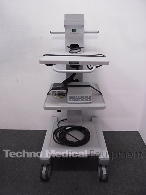
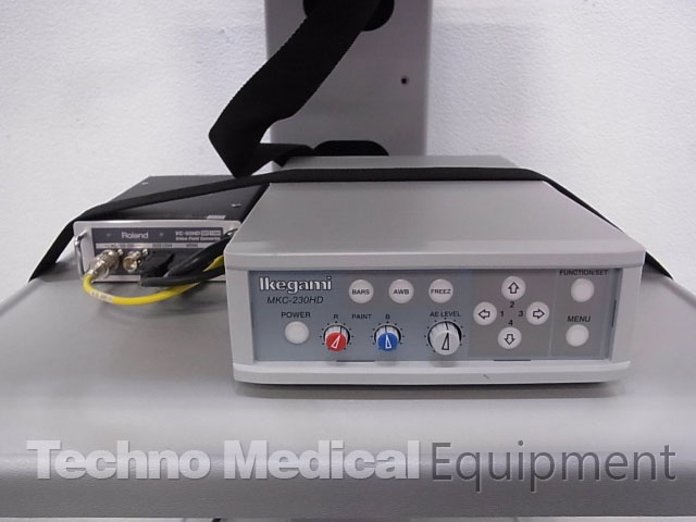
- Full high-definition camera MKC-230HD
- Video Field Converter VC-50HD
This unit comes with an SCI With Stereo Coaxial Illumination that produces a bright and uniform transillumination with less light. The depiction of the difference between the refractive power of the transparent body, creating the operative field the details of the eye. Interfaces to the CALLISTO eye OR management system
Apochromatic optics, motorized zoom system, 1:6 zoom ratio, 50 mm focusing range, 12.5x eyepiece, objective lens f=200mm.
Integrated 408 nm UV barrier filter, blue-blocking filter, retinal protection device
ZEISS OPMI Lumera i | High quality visualization
The OPMI Lumera i from ZEISS delivers high-quality visualization technology. The Stereo Coaxial Illumination (SCI) will make a difference in how well you see the details – for better decision making. See overlays of the assistance functions in the eyepiece with the External Data Injection System (EDIS).
The OPMI LUMERA family from ZEISS represents excellence in optics and illumination. The ZEISS OPMI Lumera i with ZEISS high-quality visualization technology, including Stereo Coaxial Illumination (SCI), will make a difference in how well you see the details during cataract and retina surgery.
- See the most minute structures during surgery
- Identify the details of the retina
- See overlays of the assistance functions in the eyepiece
- Manage depth of field with a push of a button
- View structures in the eye in natural colors
The 1Chip HD camera system: provides excellent visualization of natural color renditions and crisp anatomical details
Unmatched ZEISS optics: for exceptional clarity, contrast and light
RESIGHT from ZEISS: provides a clear, detailed view of the retina
Instant Red Reflex brightly: illuminates the eye – due to Stereo Coaxial Illumination (SCI) even with mature cataracts.
External Data Injection System: (EDIS) allows you to see overlays of the assistance functions in the eyepiece of your ZEISS Lumera i
Deep View depth of field: management system allows you to choose between maximum depth of field or optimum light transmission.
Cataract Surgery
For cataract surgery, SCI and CALLISTO eye® from ZEISS provide excellent anterior visualization and highly precise1,2,3 assistance functions to accelerate procedural workflow and improve surgical accuracy.
Superior red reflex: With the now firmly established Stereo Coaxial Illumination (SCI) and renowned ZEISS optics, ZEISS OPMI Lumera i brings even the most minute anatomical structures clearly into view. Its highly stable, high-contrast red reflex further enhances detail recognition.
See assistance functions in the eyepiece: Combined with the ZEISS CALLISTO eye, ZEISS OPMI Lumera i provides a series of assistance functions for performing precise1,2,3 LRI incisions, capsulorhexis, IOL centration and toric IOL alignment. All assistance functions are injected directly into the eyepiece via EDIS (External Data Injection System) as high-resolution, high-contrast images and controllable with the wireless foot control panel. This allows you to work comfortably and with full concentration without needing to look up from the eyepiece. The high-quality HD images and videos can also be displayed on the ZEISS CALLISTO eye touch screen and recorded for documentation.
| Assistance functions in the eyepiece | |||
 |
 |
 |
 |
| Incision / LRI assistant | Rhexis assistant | Z ALIGN – toric assistant | K TRACK |
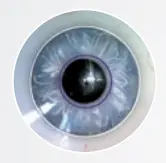
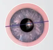
| Superimpose the exact position and size of the incisions to ensure precise 1,2,3 surgery | Superimpose the exact shape and size of the capsulorhexis and center the IOL along the optical axis of the patient‘s eye |
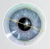
Inject reference axis and target axis in your microscope eyepiece to ensure precise1,2,3 toric IOL alignment without corneal markers |
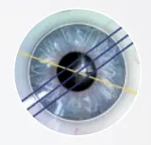
Visualize Corneal curvature in combination with a keratoscope, e. g. in corneal transplantations. |
| 1 Clinical data of Prof. Findl / Dr. Hirnschall presented at ESCRS 2013 – technically verified pre- / intraoperative matching precision ± 1.0° in mean 2 Lackerbauer, C. Modern Solutions for Refractive Cataract Surgery: CALLISTO eye. Cataract & Refractive Surgery Today. February 2013. 3 Findl, O. Complications of the CCC. Cataract & Refractive Surgery Today Europe. March 2012 |
|||
Skip unnecessary workflow steps
The ZEISS OPMI Lumera i is an integral part of the ZEISS Cataract Suite markerless – products designed to work together for precise1,2,3 and fast toric IOL alignment. You can skip manual pre- and intraoperative marking steps and manual data transfer; thereby providing a higher level of comfort for you and your patients.
Retina Surgery
ZEISS OPMI Lumera i and ZEISS RESIGHT fundus viewing systems allow you to clearly recognize the details of the retina.
ZEISS RESIGHT Family provides excellent optical quality
The non-contact fundus viewing systems provide a clear, detailed visualization of the retina. ZEISS RESIGHT 500 incorporates varioscope optics from ZEISS to keep you focused on the retina, without moving the microscope. The innovative lens turret, equipped with two aspheric lenses 128D and 60D, lets you quickly switch to a second lens and magnification. If contact is accidently made with the patient eye, the system automatically folds up. Because only sterile parts need to be changed, the optics can remain on the surgical microscope in preparation for the next patient. It’s that easy.
| Full workflow efficiency | |
 |
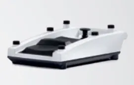 |
| Turn the world upside down | Foot control freedom |
| The Invertertube combines ZEISS optics and optical inverters into a single ergonomic design that supports a comfortable, upright working posture, without adding to stack height | Foot control freedom The foot control panel offers positioning flexibility and the ability to configure functions based on preferences |
| 4 2nd-generation optics with clearly better overall optical quality and clearly better sharpness, depth and detail recognition for use with the 60D lens – 2013 internal clinical customer survey with international key opinion leader surgeons | |
Technical specifications ZEISS OPMI Lumera i
| Technical data | |
| Surgical microscope | Apochromatic optics |
| Motorized zoom system, 1:6 zoom ratio, magnification factors γ = 0.4 to 2.4 | |
| Focusing range: 50 mm | |
| Binocular tube: 0-180° tiltable tube (optional Invertertube) | |
| Eyepieces: 10x (12.5x optional) | |
| Objective lens f = 200 mm (f = 175 mm optional) | |
| DeepView: depth of field management system | |
| Illumination | SCI: red reflex illumination and full field illumination, both are adjustable |
| Integrated 408 nm UV barrier filter | |
| Blue blocking filter | |
| Retinal protection device | |
| Fiber optic illumination | |
| Optional: fluorescence filter | |
| Light source | 12 V, 100 W halogen light source with fully automatic bulb change in case of lamp failure |
| X-Y coupling | 61 mm x 61 mm adjustment range |
| Free programmable button for starting positions of X-Y coupling, focus and zoom, light | |
| Weight of microscope | Approx. 8.5 kg (without tube, objective lens and eye pieces) |
| Suspension system | Floor stand |
| Maximum load capacity: 14 kg (complete microscope equipment, including accessories) | |
Resources: OPMI-Lumera-i_Brochure-EN.pdf