
Product Description
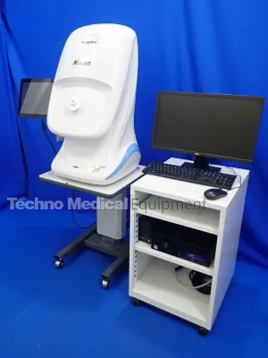
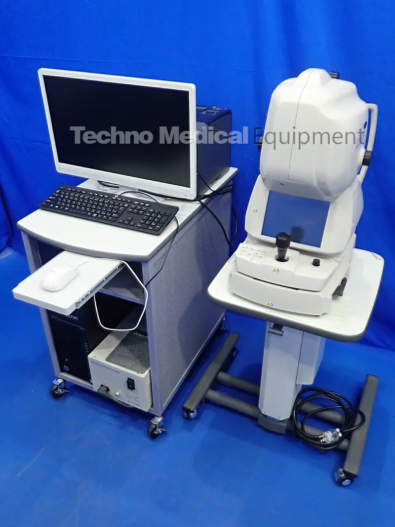
Buy Certified Used Topcon DRI Triton Plus OCT Anterior for sale, excellent patient ready condition with PC set and Anterior eye lens.
1050nm OCT for Posterior and anterior segment OCT and optional OCT Angiography imaging. Color, red free, FA and FAF photography.
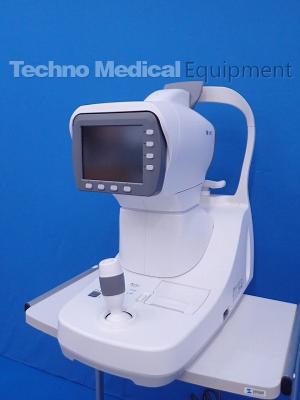
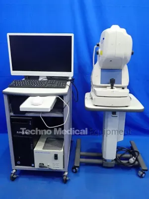
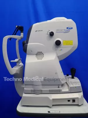
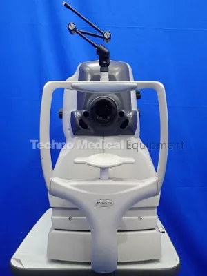
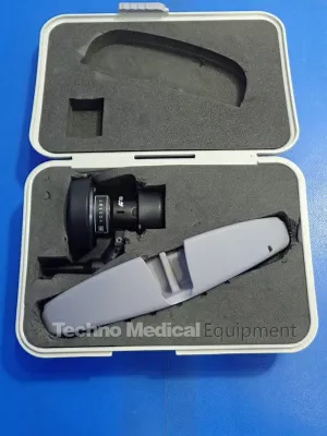
excellent condition 2017 Topcon DRI Triton Plus OCT Anterior System with Anterior eye lens unit AA, PC set, trolley, ready to work.
All items are sold in as-is condition with 12 month warranty.
Sale includes everything and ready to work:
- Topcon DRI OCT Triton Plus
- PC set with rack
- Electric optical stand
- Printer
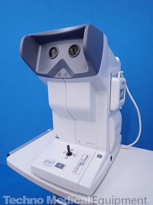
- Anterior eye lens unit AA-1
*OCT Angiography can be performed in combination with IMAGEnet 6.
The Topcon DRI OCT Triton (plus) is a swept source OCT with a non-mydriatic color fundus camera and a monochrome camera for fluorescein angiography and fundus auto fluorescence utilizing the exclusive Spaide auto fluorescence filters. Utilizing a 1,050 nm wavelength light source, and a scanning speed of 100,000 A Scans/sec, it provides uniform scanning sensitivity allowing superior visualization of the vitreous and choroid in the same scan. Invisible OCT scanning light along with high scanning speeds reduce the effect of patient eye movement and allow for more data be to collected. A 12 mm x 9 mm wide field scan along with 7 layer automated layer segmentation (including choroid) provides measurement and topographical maps of the optic nerve and macula in one scan.
The easy-to-use, intuitive IMAGEnet®6 software enables dynamic viewing of the OCT data, providing 3D, 2D and fundus images simultaneously. Pin-Point™ Registration properly indicates the location of the OCT image within the fundus image. In addition, the compare and follow up scan functions allow users to view serial exams as well as scan the exact same location of the retina. EnView software, based on en face technology, with layer flattening application allows for visualization of the various layers of the retina. Enhanced Vitreous Visualization (EVV) application allows the user to easily see the structures of the vitreous.
Key Features:
- 1 micron wavelength
allows deeper penetration into choroid and sclera
less light scattering improves results in eyes with cataracts
provides uniform sensitivity allowing superior visualization of the vitreous and choroid in the same scan
- Invisible OCT scanning light and high imaging speed of 100,000 A Scans/sec reduce the effect of eye movements and allow more data to be collected per scan
- Active Eye Tracking during capture of OCT Angiography images ensures OCT Angiography images free of motion artifiacts
- New moving image averaging improves signal-to-noise ratio giving better B scan images
- Widefield OCT, 12mm x 9 mm scan, captures the macula and disc in the same scan
- 7 layer automated segmentation (including choroid)
- Pin-Point Registration of OCT image with fundus image
- Compare function allows serial monitoring of OCT images
- Follow up scan mode allows for scanning in the exact same region of the retina
- Advanced 3D volumetric layer detection algorithms
- IMAGEnet®6 software enables dynamic viewing of 2D, 3D and fundus images simultaneously
- Embedded touch-screen for quick and easy navigation
- Automated image acquisition process (auto-focus, auto-shoot)
- Auto disc and fovea centering of OCT image
- High resolution non-mydriatic fundus camera for color, red free, stereo and panoramic fundus imaging
- Dedicated monochrome high resolution fundus camera for fluorescein angiography and fundus auto fluorescence utilizing the exclusive Spaide auto fluorescence filters
- EnView software with layer flattening application allows for visualization of the various layers of the retina
- Enhanced Vitreous Visualization (EVV) application allows the user to easily see the structures of the vitreous
- Hood report available to help quickly and easily detect glaucomatous damage.
TOPCON DRI OCT Triton Plus Manufacturer Technical data
| Observation & Photography of Fundus Image | Photography Type: Color, FA*, FAF*, Red-free** |
| Picture Angle: 45° Equivalent 30° (Digital Zoom ) |
|
| Operating Distance: 34.8mm | |
| Photographable Diameter of Pupil: Normal:φ4.0mm or more Small pupil diameter: φ3.3mm or more |
|
| Observation & Photography of Fundus Tomogram | Scanning Range (on fundus): Horizontal Within 3 to 12mm Vertical Within 3 to 12mm |
| Scan Pattern: 3D scan Linear scan (Line-scan/Cross-scan/Radial-scan) |
|
| Scan Speed: 100,000 A-Scans per second | |
| Lateral Resolution: 20μm | |
| In-depth Resolution: Digital: 2.6μm Optical function: 8μm |
|
| Photographable Diameter of Pupil: φ2.5mm or more | |
| Observation & Photography of Fundus Image / Fundus Tomogram | Fixation target; Internal fixation target : Dot matrix type organic EL The display position can be changed and adjusted. The displaying method can be changed. Peripheral fixation target : This is displayed according to the internal fixation target displayed position. External fixation target |
| Observation & photography of anterior segment*** | Photography type: IR |
| Operating distance: 17mm | |
| Observation & photography of anterior segment tomogram*** | Operating distance: 17mm |
| Scan range (on cornea): Horizontal Within 3 to 16mm Vertical Within 3 to 16mm |
|
| Scan pattern: 3D scan Linear scan (Line-scan/Radial-scan) |
|
| Scan speed: 100,000 A-Scans per second | |
| Fixation target: Internal fixation target External fixation target |
|
| Electric Rating | Power Source: Voltage: 100-240V Frequency: 50-60Hz |
| Power input: 250VA | |
| Dimensions / Weight | Dimensions: 320-359 mm(W) X 523-554 mm(D) X 560-590 mm(H) |
| Weight: 21.8kg (DRI OCT Triton) 23.8kg(DRI OCT Triton plus) |
Resources; TOPCON DRI OCT Triton Plus Brochure
Related Equipment
You may also be interested in the following medical equipment.
Zeiss OPMI VISU 200 S88 Microscope with Integrated Assistant Scope