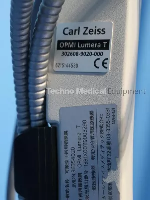
Product Description
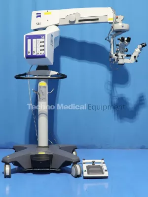
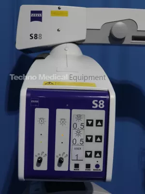
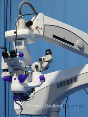
Buy Certified Used Zeiss OPMI Lumera T S88 System with Integrated Assistant Scope, Foot Switch, ready to work.
Performed all upgraded 2024, compensated and calibrated in compliance with ZEISS working standards, sold in as-is condition with 24 month warranty.
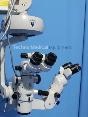
The Zeiss Lumera T on S88 Stand is an outstanding microscope designed for a full suite of ophthalmic procedures, excellent for the University Medical Center and specialists in vitreo retinal procedures. If you are looking to achieve the best in red reflex technology, look no further than the Lumera T S88 ophthalmic surgical microscope. With the introduction of Lumera Stereo Coaxial Illumination you see more contrast with this brilliant, stable red reflex – even in decentered or strongly pigmented eyes. The Lumera T S88 floor stand provides superior surgeon control from the ergonomic hand grips to release the magnetic clutch brakes moving the microscope across the surgical field with minimal effort. Program the Lumera T S88 with up to 9 different users with preset motorized speeds, illumination intensity and foot pedal controls.
Specs:
• Stereo Coaxial Illumination (SCI) incorporates illumination technology that optimizes red reflex with heavy detail recognition
• Integrated assistant's microscope with independent focusing and zoom
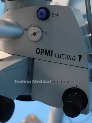
• DeepView, integrated depth-of-field management system, permits optimization of the microscope image to depth of field or light transmission
• Superlux Eye xenon illumination system offers surgeons white, natural, high contrast image of surgical field
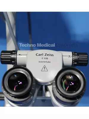
• Binocular Tube: Apochromatic optics, invertertube
• Eyepiece: 10x
• Magnification Changes: Motorized zoom system, 1:6 zoom ratio
• Total Magnification: 0.4 - 2.4 x
• Red reflex illumination and surrounding field illumination, both dimmable
• Superlux Eye Xenon Light Source
• HaMode and blue blocking filters
• Base Size 31.69 in. x 31.69 in. (805 mm x 805 mm); Height 74.02 in. (1880 mm)
Accessories:
- Assistant Binoculars
- Foot Switch
Features
Visualization of the Red Reflex
The Stereo Coaxial Illumination (SCI) allows you to benefit from the detail recognition, high-contrast brilliance and stability of the red reflex – even with strongly pigmented, decentered and ametropic eyes. This technology enables you to see all the details of a patient’s eye.
Effortless positioning
The magnetic brakes make positioning the surgical microscope very simple. When the brakes are released, the system smoothly glides into a new position; when locked, the surgical microscope remains firmly in place.
Independent second view
This OPMI Lumera® T surgical microscope is equipped with a completely integrated assistant’s microscope. The second surgeon selects the focus and magnification independently of the main surgeon, thus enabling active assistance.
Natural color impression
The integrated Superlux® Eye xenon illumination allows you to see the anatomic structure of the eye in its natural colors and highly accurate detail. The use of the HaMode™ filter allows surgeons who prefer halogen to quickly switch to a light spectrum equivalent to halogen. This is particularly beneficial when several surgeons with different preferences regarding the light source use one system.
Deep View the integrated depth-of-field management system, permits optimization of the microscope image to depth of field or light transmission.
Superlux Eye xenon illumination offers surgeons a whiter, more natural, higher contrast image of the surgical field than standard halogen illumination.
HaMode filter for Superlux Eye xenon illumination system, generates halogen-like light Ideal for hospitals where some surgeons prefer xenon, others halogen illumination.
SCI (Stereo Coaxial Illumination) provides constant brilliance and brightness: every detail of the patient’s eye becomes visible.
Apochromatic optics with high light transmission, result in maximum image quality for operations and documentation.
ZEISS OPMI LUMERA T Manufacturer Technical data
| Surgical microscope | Apochromatic optics |
| Motorized zoom system, 1:6 zoom ratio, magnification factors = 0.4 to 2.4 | |
| Focusing range: 50 mm | |
| Binocular tube: Invertertube® (optional 0-180° tiltable tube) | |
| Eyepieces: 10x (12.5x optional) | |
| Objective lens f = 200 mm (f = 175 mm optional) | |
| DeepView: depth of field management system | |
| Integrated assistant’s microscope | |
| Fully Stereoscopic | |
| Illumination | SCI: red reflex illumination and surrounding field illumination, both dimmable, patent pending |
| Retinal protection device | |
| Fiber optic illumination | |
| Light source | Superlux Eye xenon Light Source with manual bulb change |
| HaMode filter | |
| Optional: 12V, 100W halogen Light Source with fully automatic bulb change in case of lamp failure | |
| Option: dual halogen Light Source | |
| Optional: xenon / halogen combi Light Source | |
| Integrated 408 nm UV barrier filter | |
| Blue-blocking filter | |
| Optional: fluorescence filter | |
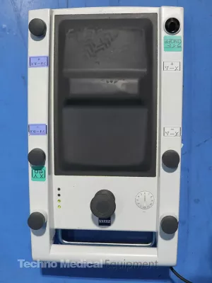
| X-Y coupling | 40 mm x 40 mm adjustment range |
| Button for starting positions of the X-Y coupling and focus | |
| Weight | 13.7 kg (30.2 lb) (with Invertertube, integrated assistant’s microscope, objective lens and eyepieces) |
| Suspension system | S88 floor stand |
| Maximum load: 20 kg (44.1 lb) (complete microscope equipment, including accessories) |
Resources; Carl Zeiss OPMI Lumera T Brochure
Related Equipment
You may also be interested in the following medical equipment.
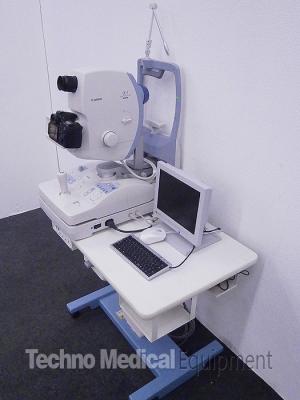
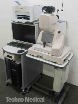
Canon CX-1 Hybrid Digital Retinal Camera
Zeiss OPMI Lumera 700 Microscope with Resight 700